38 compound microscope diagram
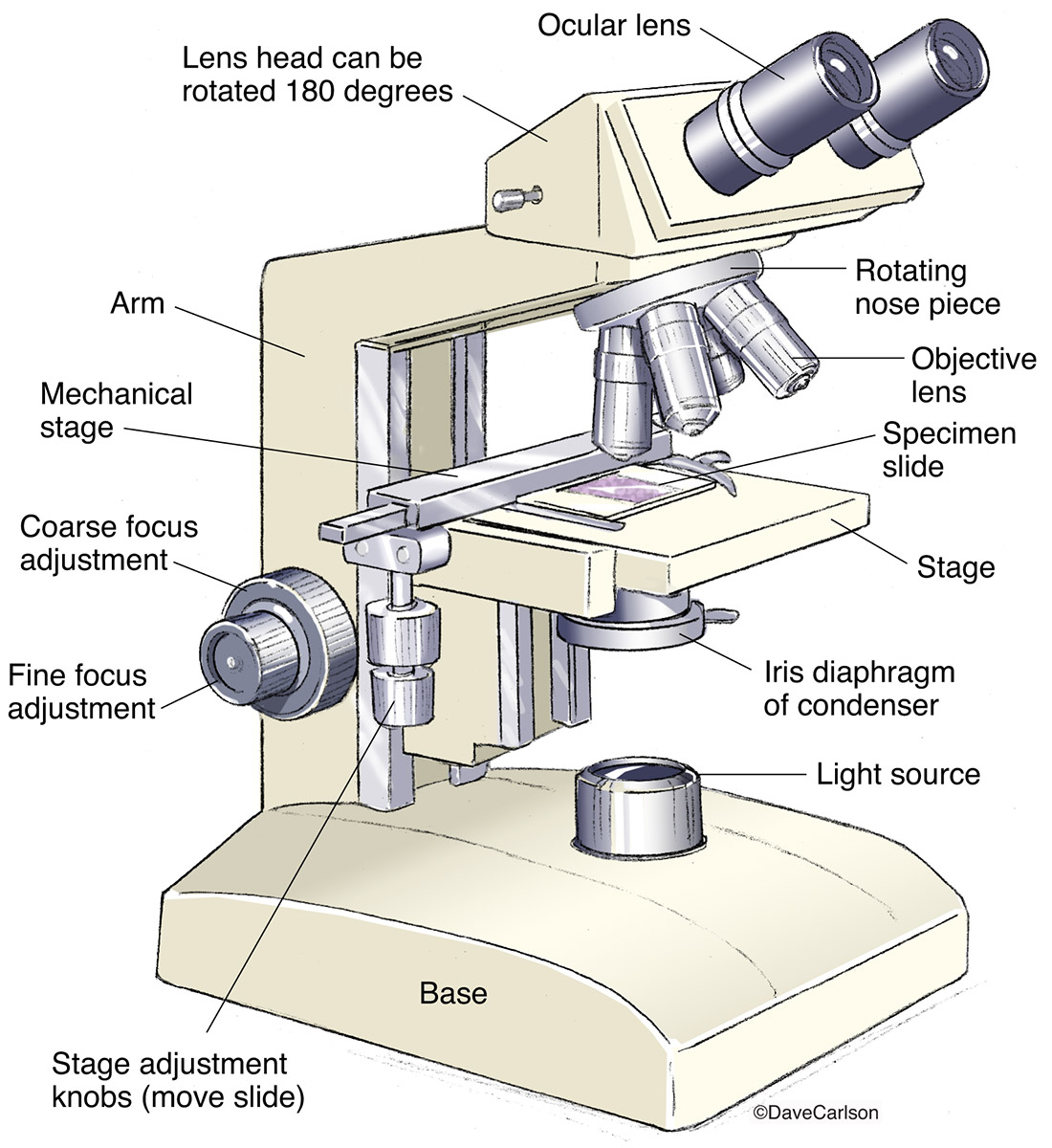
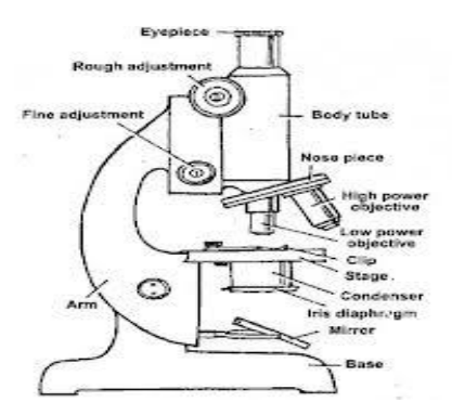
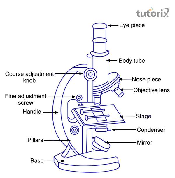
Compound Microscope - Types, Parts, Diagram, Functions and ... Mar 7, 2022 · A compound microscope has two convex lenses; an objective lens and eye piece. The objective lens is placed towards the object and the eyepiece is the lens towards our eye. Both eyepiece and objective lenses have a short focal length and fitted at the free ends of two sliding tubes. (4, 5, and 6) Compound microscope parts and magnification Compound Microscope Parts – Labeled Diagram and their ... There are three major structural parts of a compound microscope. The head includes the upper part of the microscope, which houses the most critical optical components, and the eyepiece tube of the microscope. The base acts as the foundation of microscopes and houses the illuminator. The arm connects between the base and the head parts.
Microscope Parts and Functions With Labeled Diagram and ... Learning to use and adjust your compound microscope is the next important step. It's also imperative to know and understand the best practices of cleaning your microscope . The microscope parts work together in hospitals and in forensic labs, for scientists and students, bacteriologists and biologists so that they may view bacteria, plant and animal cells and tissues, and various microorganisms the world over.

Compound microscope diagram
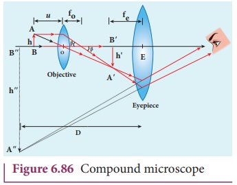
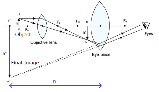
Compound Microscope: Diagram, Parts, Working & Magnification ... Working of compound microscope As shown in figure the lens near the object is called the objective which forms a real, inverted, magnified image of the object. This image serves as the object for the second lens. The lens which is used to view the final image is called an eyepiece, which functions like a simple microscope or magnifier. Compound Microscope Parts, Functions, and Labeled Diagram Nov 18, 2020 · The individual parts of a compound microscope can vary heavily depending on the configuration & applications that the scope is being used for. Common compound microscope parts include: Compound Microscope Definitions for Labels Eyepiece (ocular lens) with or without Pointer: The part that is looked through at the top of the compound microscope. Eyepieces typically have a magnification between 5x & 30x. Parts of a microscope with functions and labeled diagram Sep 17, 2022 · Figure: Diagram of parts of a microscope There are three structural parts of the microscope i.e. head, base, and arm. Head – This is also known as the body. It carries the optical parts in the upper part of the microscope. Base – It acts as microscopes support. It also carries microscopic illuminators.
Compound microscope diagram. Diagram of a Compound Microscope - Biology Discussion Diagram of a Compound Microscope Article Shared by ADVERTISEMENTS: In this article we will discuss about:- 1. Essential Parts of Compound Microscope 2. Magnification of the Image of the Object by Compound Microscope 3. Resolution Power 4. Method for Studying Microbes 5. Measurement of the Size of Objects. Essential Parts of Compound Microscope: Parts of a microscope with functions and labeled diagram Sep 17, 2022 · Figure: Diagram of parts of a microscope There are three structural parts of the microscope i.e. head, base, and arm. Head – This is also known as the body. It carries the optical parts in the upper part of the microscope. Base – It acts as microscopes support. It also carries microscopic illuminators. Compound Microscope Parts, Functions, and Labeled Diagram Nov 18, 2020 · The individual parts of a compound microscope can vary heavily depending on the configuration & applications that the scope is being used for. Common compound microscope parts include: Compound Microscope Definitions for Labels Eyepiece (ocular lens) with or without Pointer: The part that is looked through at the top of the compound microscope. Eyepieces typically have a magnification between 5x & 30x. Compound Microscope: Diagram, Parts, Working & Magnification ... Working of compound microscope As shown in figure the lens near the object is called the objective which forms a real, inverted, magnified image of the object. This image serves as the object for the second lens. The lens which is used to view the final image is called an eyepiece, which functions like a simple microscope or magnifier.
Optical Instruments: Compound Microscope and its Magnification

Draw a labelled ray diagram of compound microscope and derive ...

A schematic of a basic compound microscope. | Download ...

Simple doodles, Microscopic images, Microscope parts

JaypeeDigital | eBook Reader

Binocular Microscope Anatomy - Parts and Functions with a ...

Compound Microscope Parts, Functions, and Labeled Diagram ...
Compound microscope - Optical Instruments

Lab Essentials | Compound Microscope Basics – LabEssentials, Inc.
Anatomy of a Microscope | Microscopy Primer | Olympus LS

easy compound microscope diagram - Clip Art Library
a)Draw a ray diagram of a compound microscope for the final ...

Can someone can send me diagram of this compound microscope ...
Compound Microscope | Image License | Carlson Stock Art

Simple Microscope Diagram, Formula, Definition, Discoverd by

a Draw a ray diagram for the formation of image by a compound ...

How to draw ray diagram of compound microscope

Compound Microscope: Basics, Functionality, and Uses ...

Draw A Ray Diagram Of A Compound Microscope - Diagram ...
Draw a neat labelled diagram of a compound microscope and ...

Draw a neat labelled diagram of a compound microscope. Derive ...

a) Explain the working of a compound microscope with the help ...

Compound Microscope: Definition, Parts, Application, Working ...
Compound Microscope.

Compound Microscope Diagram | Quizlet

KOPAL Classes - Compound Microscope Ray Diagram | Facebook
Difference Between Simple and Compound Microscope

Compound Microscope: Diagram, Parts, Working & Magnification ...

How to draw compound of Microscope easily - step by step

The Compound Microscope Diagram | Quizlet

Living Environment Course

Compound Microscope Parts, Functions, and Labeled Diagram ...
Compound Microscope, Ray Diagram Mistakes. | Physics Forums

Draw a Ray Diagram Showing Image Formation in a Compound ...

Compound and Stereo- microscopes - Microscopes 4 Schools

Compound Microscope Parts, Functions, and Labeled Diagram ...

Parts of a microscope with functions and labeled diagram

Microscope | Types, Parts, History, Diagram, & Facts | Britannica
Post a Comment for "38 compound microscope diagram"