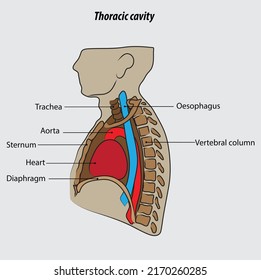
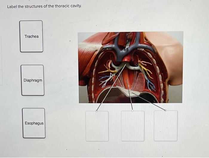
43 label the structures of the thoracic cavity.
681 Thoracic cavity Images, Stock Photos & Vectors - Shutterstock 681 thoracic cavity stock photos, vectors, and illustrations are available royalty-free. See thoracic cavity stock video clips. Image type. Quiz & Worksheet - Pleural Cavities and Pleural Membranes - Study.com Topics assessed in this quiz/worksheet combo include: The relationship between the thoracic and abdominal cavities. The names of membranes lining the thoracic cavity and lungs. Pressure ...
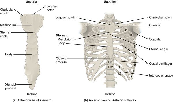
Bones of the Thoracic Wall - 3D Models, Video Tutorials & Notes ... You've got structures like the aorta and the inferior vena cava as well as the oesophagus, which pass through the diaphragm. In terms of the bony framework which makes up the thorax, you've got the 12 th thoracic vertebra at the back, which make up the posterior part of the thorax. And then you've got the ribs which form the lateral part.
Label the structures of the thoracic cavity.
Anatomy, Thorax - StatPearls - NCBI Bookshelf The thoracic cavity is found deep to the thoracic wall, superior to the diaphragm, and inferior to the root of the neck (thoracic aperture). The thoracic cavity contains organs and tissues that function in the respiratory (lungs, bronchi, trachea, pleura), cardiovascular (heart, pericardium, great vessels, lymphatics), nervous (vagus nerve ... Ventral Body Cavity | Subdivisions, Organs, & Diagram - Video & Lesson ... The thoracic cavity contains the lungs, each enclosed by a pleural cavity; the heart, enclosed by the pericardial cavity; the trachea; and the esophagus. Abdominopelvic Cavity The abdominopelvic... Thoracic cavity | Whitman College Thoracic cavity Section Navigation In this photo, we can see the heart contained within in its transparent pericardial membrane. Also found inside the thoracic cavity are the right and left lungs, which are on either side of the heart. Also note the thymus gland, which in many young mammals can be found in the throat and the thoracic cavity.
Label the structures of the thoracic cavity.. Thoracic cavity - Wikipedia Structures within the thoracic cavity include: structures of the cardiovascular system, including the heart and great vessels, which include the thoracic aorta, the pulmonary artery and all its branches, the superior and inferior vena cava, the pulmonary veins, and the azygos vein Body Cavities and Organs | Biology Dictionary This cavity is the true coelom, as it forms during human embryogenesis from the mesoderm. At first it is a single cavity. It then gets subdivided several times, into smaller cavities separated by muscles, bones, and thin tissues. The first subdivision is the diaphragm muscle, which divides the abdominopelvic cavity from the thoracic cavity ... Body Cavities and Membranes - Anatomy and Physiology Notes The ventral body cavity is the larger cavity located toward the front of the body, and it contains our visceral organs (or guts!). Remember: ventral contains the viscera! The ventral cavity can also be divided into two main parts: the thoracic cavity and abdominopelvic cavity, which are separated by the diaphragm. Thoracic Cage Labeling Quiz - PurposeGames.com This is an online quiz called Thoracic Cage Labeling. There is a printable worksheet available for download here so you can take the quiz with pen and paper. Your Skills & Rank. Total Points. 0. Get started! Today's Rank--0. Today 's Points. One of us! Game Points. 13. You need to get 100% to score the 13 points available.
Body Cavities and Membranes: Labeled Diagram, Definitions - EZmed As shown in the flow chart above, the thoracic cavity can be subdivided into 2 main parts: Pleural Cavities. Mediastinum. Let's look at the pleural cavities first followed by the mediastinum. Features of the Pleural Cavities. Location: Right and Left Thoracic Cavity. Surrounds: Lungs. Fluid: Pleural Fluid. Membrane: Pleura (Visceral Pleura and Parietal Pleura) Thoracic Cavity - Introduction, Structure, Organs, and FAQs - VEDANTU Structures within the thoracic cavity include: Oesophagus of the digestive system Thymus gland Vagus nerve and parasympathetic veins. Diaphragm, trachea, bronchi, lungs. The heart The superior and inferior vena cava. Pulmonary vein and artery. The thoracic cavity diagram is drawn below: I m a g e w i l l b e u p l o a d e d s o o n Pleural Membrane thoracic cavity | Description, Anatomy, & Physiology | Britannica thoracic cavity, also called chest cavity, the second largest hollow space of the body. It is enclosed by the ribs, the vertebral column, and the sternum, or breastbone, and is separated from the abdominal cavity (the body's largest hollow space) by a muscular and membranous partition, the diaphragm. Thoracic and mediastinal lymph nodes and lymphatics | Kenhub The thorax is the region of the body extending from the base of the neck and thoracic inlet (the latter being at the supraclavicular fossae) to the diaphragm (marked anteriorly by the xiphisternal joint). Within the thoracic cavity is the mediastinum. The mediastinum is the region of the thorax between the lungs.
Label Thoraic Cavity 2.png - l View site information l... View Homework Help - Label Thoraic Cavity 2.png from BIO 141 at Northern Virginia Community College. l View site information l Label the structures of the thoracic Study Resources Main Menu Thorax: Anatomy, wall, cavity, organs & neurovasculature | Kenhub It is made up of the sternum, twelve pairs of ribs, twelve thoracic vertebrae, and interconnecting joints. The main thoracic joints include the intervertebral discs, costovertebral, sternocostal, sternoclavicular, costochondral, and interchondral joints. Running between every two adjacent ribs are anatomical spaces called intercostal spaces. Body Cavities and Organs with Labeled Diagram - AnatomyLearner The thoracic cavity of an animal is cone-shaped and laterally compressed. In addition, the abdominal cavity of the animal is the largest cavity that extends from the diaphragm to the pelvic inlet. ... Now, you might know the different structures of the animal nasal cavity. Hey, don't forget to check the frequently asked question and labelled ... Organs in the Thoracic Cavity - Bodytomy The ribs, vertebral column, muscles, connective tissues, and the sternum (breast bone) enclose this cavity. The thoracic cavity is lined by a serous membrane that exudes a thin fluid (serum). The chest membrane, also known as parietal pleura, extends further to cover the lungs. This part of the membrane is known as the visceral pleura.
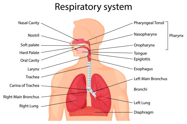
63 Drawing Of The Diagram Of The Respiratory System With ...
The Thoracic Cavity - Human Anatomy The Thoracic Cavity The heart and lungs are situated in the thorax, the walls of which afford them protection. The heart lies between the two lungs, and is enclosed within a fibrous bag, the pericardium, while each lung is invested by a serous membrane, the pleura.

Body Organization. - ppt download
Anatomy Chapter 1: Labeling Thoracic Cavity Diagram | Quizlet The cavities surrounding each lung parietal pleura The aspect of the pleura that does not touch the surface of the lung visceral pleura The aspect of the pleura that covers the external surface of the lung The thoracic cavity can be subdivided into... 1. mediastinum 2. left and right pleural cavities 3. pericardial cavity
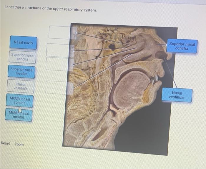
Answered: Label these structures of the upper… | bartleby
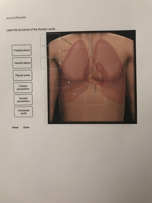
Solved Award: 0.76 points Label the structures of the - Chegg Question: Award: 0.76 points Label the structures of the thoracic cavity. Parietal pleura Visceral pleura Pleural cavity Parietal pericardium Visceral pericardium Pericardial cavity Reset Zoom This problem has been solved! See the answer Show transcribed image text Expert Answer 100% (19 ratings)

AHCDW15Notes6.pdf - 6. Award: 1.00 point Problems? Adjust ...
Thoracic Cavity Game - purposegames.com This is an online quiz called Thoracic Cavity Game. There is a printable worksheet available for download here so you can take the quiz with pen and paper. Your Skills & Rank. Total Points. 0. Get started! Today's Rank--0. Today 's Points. One of us! Game Points. 9. You need to get 100% to score the 9 points available.

The thoracic cavity | Thoracic cavity, Respiratory system ...
Label the thoracic cavities.docx - Label the cavities... In the figure above - locate the thoracic cavity. Labelthe structure that separates the thoracic cavity from the abdominopelvic cavity Notice the 4 colors of the thoracic cavity. There are two purple cavities within the thoracic cavity. Labelthem. Identifythe two / three primary structures that lie within the purple cavity. Notice the green cavity.
thoracic cavity | Description, Anatomy, & Physiology | Britannica
Thoracic Cavity - Anatomy | Organs | Functions | 8 Types of Cavities The thoracic cavity contains the center and lungs, each of that is perpetually acquiring and increasing. The ribs within the thoracic cavity serve each as protection and support, permitting the lungs to expand and contract while not running the chance of swing itself into a dangerous scenario, as well as even external threats. The abdominal contents, opposingly, are a unit of additional muscular and fewer vulnerable to injury and don't would like such excessive protection.
683 Thoracic cavity Images, Stock Photos & Vectors | Shutterstock
Unit 1 Lab Homework Flashcards | Quizlet Label the structures of the thoracic cavity. Left Down: Parietal Pleura Pleural Cavity Visceral Pleura Visceral Pericardium Pericardial Cavity Parietal Pericardium Label the directional terms based on the arrows. Left: Medial Proximal Right: Lateral Distal Label the body planes. Left: Coronal Plane Oblique Plane Right: Midsagittal Plane

Human Skeleton System Thoracic Skeleton with Label Design ...
Thoracic Cavity - Definition & Organs of Chest Cavity - Biology Dictionary The thoracic cavity is actually composed of three spaces each lined with mesothelium, a special film-like tissue that separates vital organs. The pleural cavities surround the lungs, while the pericardial cavity surrounds and protects the heart. These tissues in the thoracic cavity can be seen in the image below.
Solved] Please help me label the Cavities | Course Hero
points Label the structures of the thoracic cavity. Parietal pleura ... Classtheta 19 subscribers Question: Award: 0.76 points Label the structures of the thoracic cavity. Parietal pleura Visceral pleura Pleural cavity Parietal pericardium Visceral pericardium...
Introduction to Human Anatomy and Physiology Anatomy the
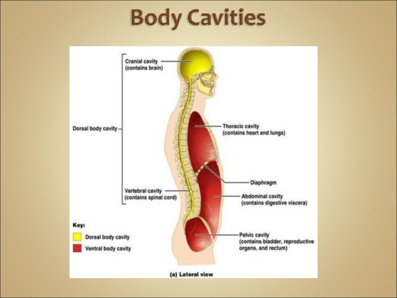
Body cavities and membranes : Anatomy & Physiology The cranial cavity is the area within the skull and encloses the brain. The spinal (vertebral) cavity encases the vertebral column and spinal cord. Ventral Body cavity. Like the dorsal cavity, the ventral cavity has two subdivisions. The superior division is called the thoracic cavity. The thoracic cavity is surrounded by the ribs and muscles ...
Imaging of the Lungs and Pleura | Concise Medical Knowledge
Thoracic cavity | Whitman College Thoracic cavity Section Navigation In this photo, we can see the heart contained within in its transparent pericardial membrane. Also found inside the thoracic cavity are the right and left lungs, which are on either side of the heart. Also note the thymus gland, which in many young mammals can be found in the throat and the thoracic cavity.

02.4 Unit 17 Pre-Lab Exercise 17-2.doc - Pre-Lab Exercise 17 ...
Ventral Body Cavity | Subdivisions, Organs, & Diagram - Video & Lesson ... The thoracic cavity contains the lungs, each enclosed by a pleural cavity; the heart, enclosed by the pericardial cavity; the trachea; and the esophagus. Abdominopelvic Cavity The abdominopelvic...

Solved Correctly label the following structures related to ...
Anatomy, Thorax - StatPearls - NCBI Bookshelf The thoracic cavity is found deep to the thoracic wall, superior to the diaphragm, and inferior to the root of the neck (thoracic aperture). The thoracic cavity contains organs and tissues that function in the respiratory (lungs, bronchi, trachea, pleura), cardiovascular (heart, pericardium, great vessels, lymphatics), nervous (vagus nerve ...

Summary Sheet 1 Questions - Human Anatomy and Physiology ...

Label Thoraic Cavity 2.png - l View site information l Label ...
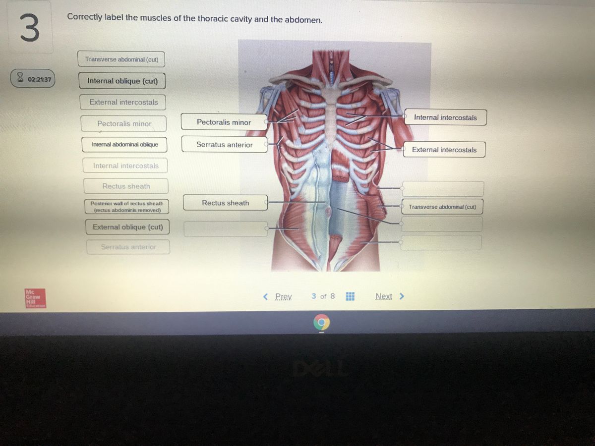
Answered: Correctly label the muscles of the… | bartleby

Thoracic Cavity Labeling Diagram | Quizlet

Human Skeleton System Thoracic Skeleton with Label Design ...

Pin on School Stuff

Thoracic cavity - Knowledge @ AMBOSS

Thoracic Cage Labeling Quiz
Solved Label the structures of the thoracic cavity. Trachea ...

Label Thoraic Cavity 2.png - l View site information l Label ...
Ribs - Physiopedia
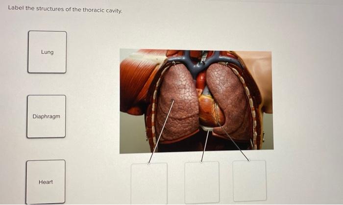
Solved Label the structures of the thoracic cavity. Lung ...
Solved Award: 0.76 points Label the structures of the | Chegg.com

Thoracic cavity - Wikipedia

The thoracic cage is made up of bones and cartilage along ...

Body cavity - Wikipedia

Anatomical structure of the thoracic cavity. | Download ...
Introduction to Anatomy, Chapter 1

Thoracic Skeleton of Human Skeleton System Anatomy with ...
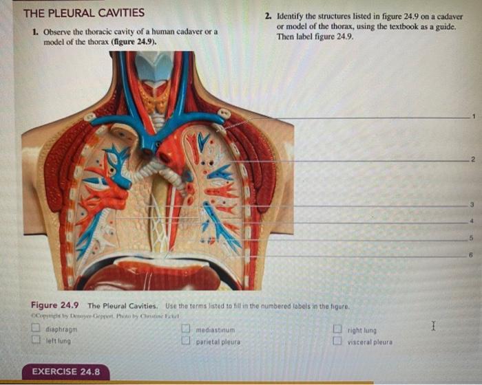
Solved THE PLEURAL CAVITIES 1. Observe the thoracic cavity ...
Pleural cavity: Anatomy, location, function | Kenhub

Professional Medical Anatomy of Human Organ System Trunk Thoracic Cavity Structure Model of The Internal Organs

Ultrasound of the Lung: Clinical Applications - ScienceDirect

Anatomy of the thoracic skeleton. Netter illustration used ...

Anatomy of the Thoracic Wall, Pulmonary Cavities, and ...
Thoracic cage: Anatomy and clinical notes | Kenhub
Torsos

AP 2 (part 3) Flashcards | Quizlet

Human Skeleton System Thoracic Skeleton with Label Design ...

Thoracic Cavity Diagram | Quizlet
Post a Comment for "43 label the structures of the thoracic cavity."