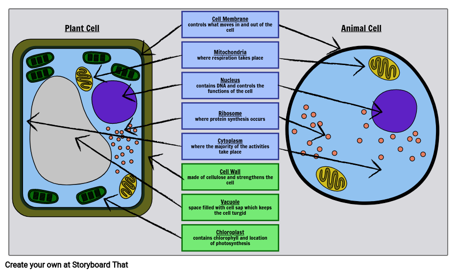
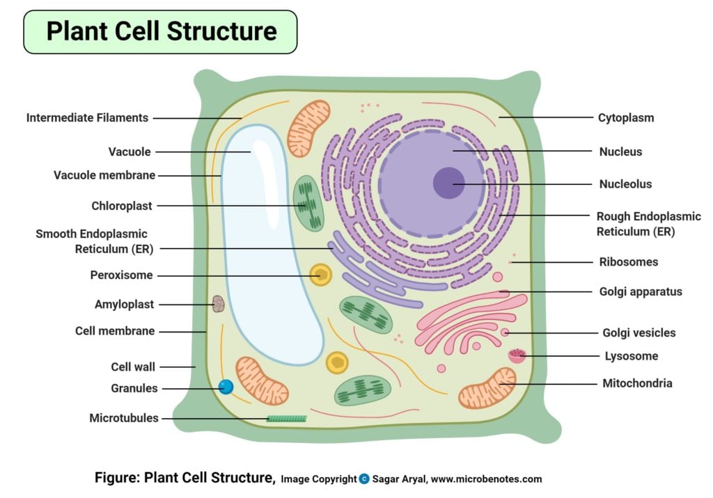
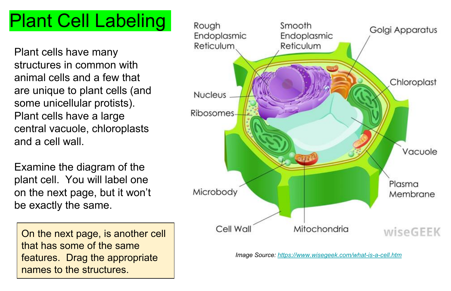
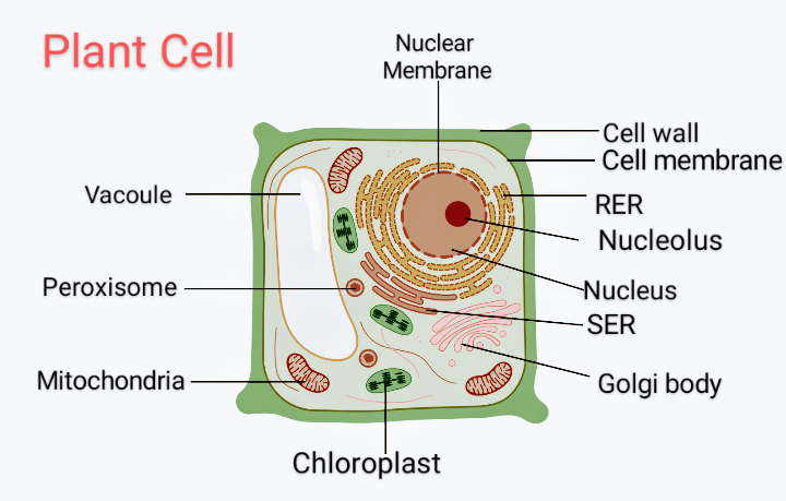
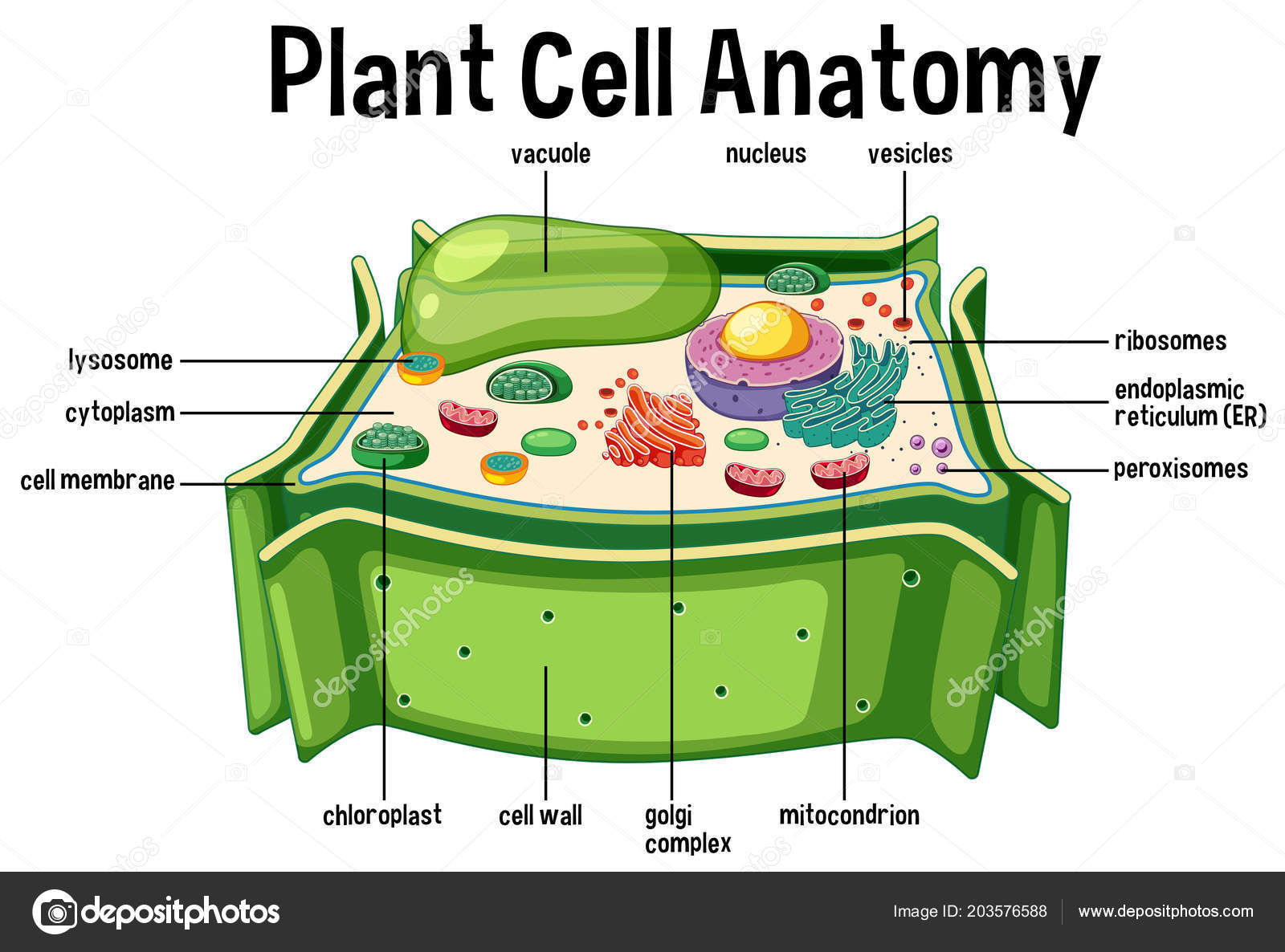
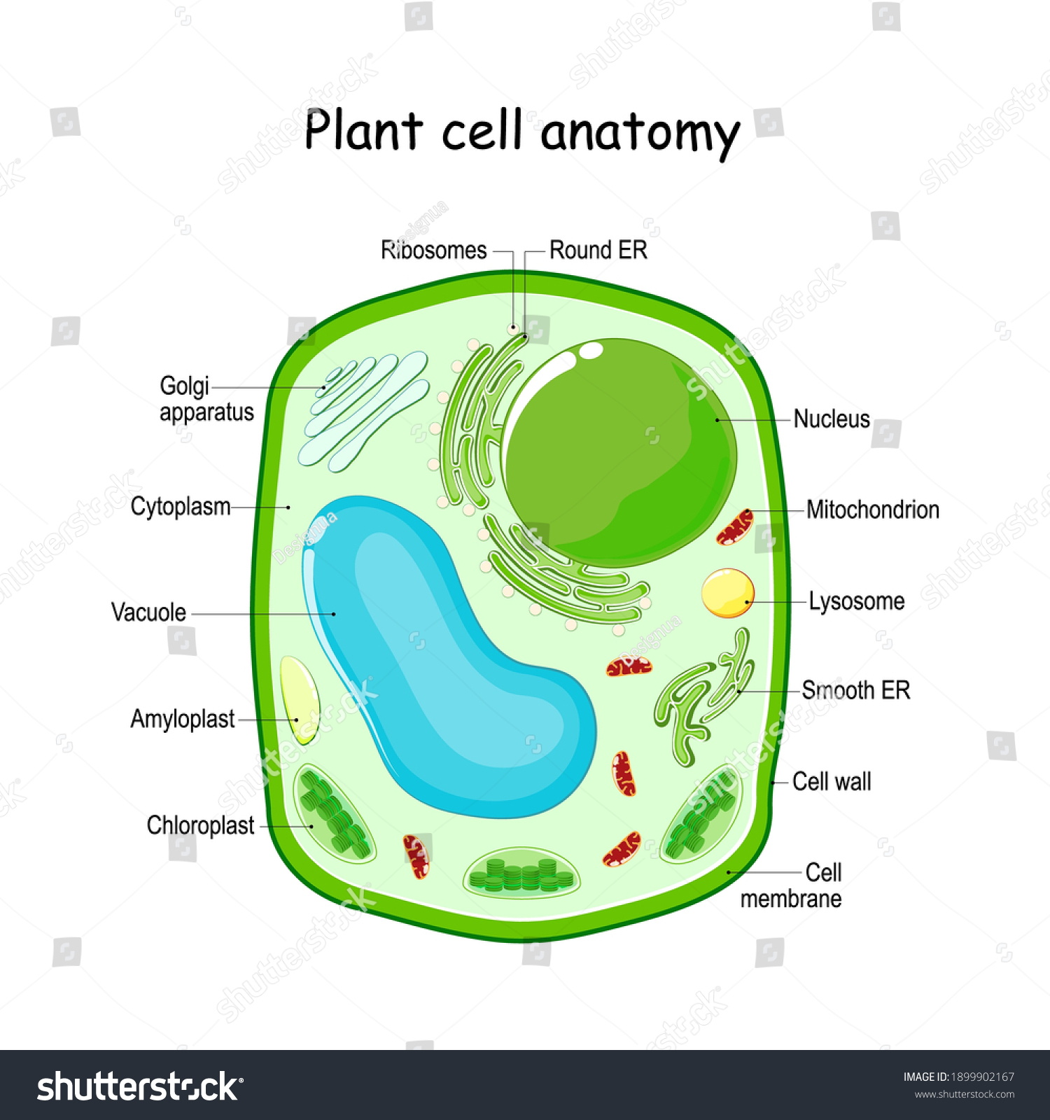
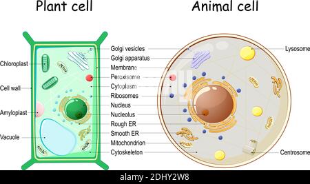
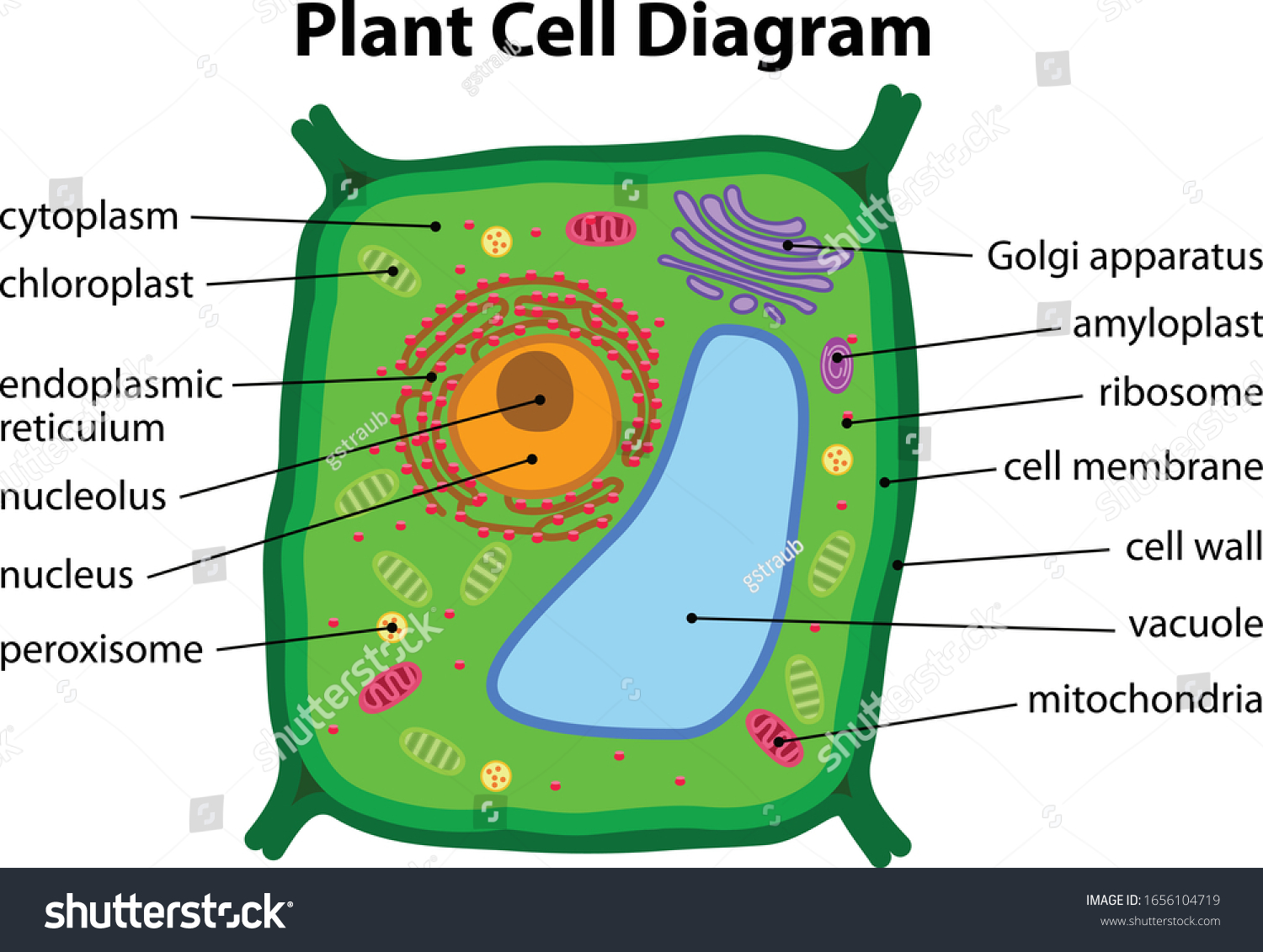
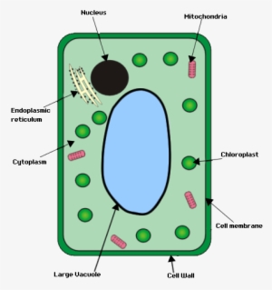
39 labeled plant cell diagram
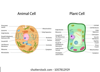
Plant Cell - The Definitive Guide | Biology Dictionary Jan 15, 2021 · A diagram of a plant cell with the organelles labeled The plant cell has many different features that allow it to carry out its functions. Each of these structures, called organelles, carry out a specialized role. Compound Microscope- Definition, Labeled Diagram, Principle, … 03.04.2022 · Light Microscope- Definition, Principle, Types, Parts, Labeled Diagram, Magnification; Amazing 27 Things Under The Microscope With Diagrams; 22 Types of Spectroscopy with Definition, Principle, Steps, Uses; Plant Cell- Definition, Structure, Parts, Functions, Labeled Diagram; Advantages. Simplicity and its convenience. A compound light …
Compound Microscope- Definition, Labeled Diagram, Principle ... Apr 03, 2022 · Occasionally very high magnification it required (e.g. for observing bacterial cell). In that case, an oil immersion objective lens (usually 100X) is employed. The common light microscope is also called a bright-field microscope because the image is produced amidst a brightly illuminated field.

Labeled plant cell diagram
Microscope Parts and Functions Microscope Parts and Functions With Labeled Diagram and Functions How does a Compound Microscope Work?. Before exploring microscope parts and functions, you should probably understand that the compound light microscope is more complicated than just a microscope with more than one lens.. First, the purpose of a microscope is to magnify a small object or to … Microscope, Microscope Parts, Labeled Diagram, and Functions 19.01.2022 · Microscope cell staining is a technique used to improve the visibility of cells and cell parts under a microscope. A nucleus or a cell wall can be seen more clearly by using different stains. 2. Iodine, crystal violet, and methylene blue are examples of simple stains. 3. Make a wet or dry mount with a coverslip. 4. A student constructs a Venn diagram to compare the ... - BRAINLY Nov 13, 2020 · Venn Diagram of Plant and Animal Cells A Venn Diagram is shown. One circle is labeled Animal only, the other circle is labeled plant only, and the overlapping section is labeled both. Which organelle should be listed under “Animal Only” in the diagram? nucleus centriole ribosome cell wall" Is B. Centriole
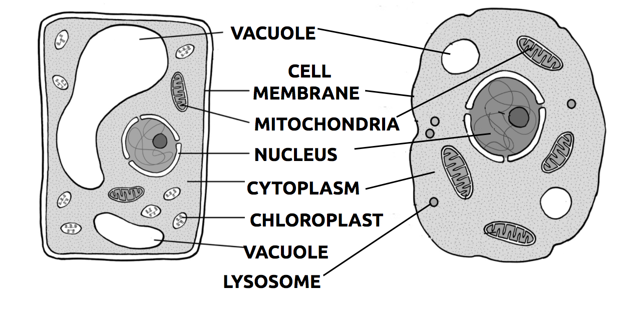
Labeled plant cell diagram. Plant Cell- Definition, Structure, Parts, Functions, Labeled Diagram 16.02.2022 · Figure: Labeled diagram of plant cell, created with biorender.com. The typical characteristics that define the plant cell include cellulose, hemicellulose and pectin, plastids which play a major role in photosynthesis and storage of starch, large vacuoles responsible for regulating the cell turgor pressure. Microscope, Microscope Parts, Labeled Diagram, and Functions Jan 19, 2022 · Microscope cell staining is a technique used to improve the visibility of cells and cell parts under a microscope. A nucleus or a cell wall can be seen more clearly by using different stains. 2. Iodine, crystal violet, and methylene blue are examples of simple stains. 3. Make a wet or dry mount with a coverslip. 4. Transcriptional landscape of rice roots at the single-cell resolution 01.03.2021 · Venn diagram analyses showed that more than 80% of expressed genes were shared between Nip and 93-11 in all big cell-type clusters, including cortex, endodermis, epidermis, and stele. For the cultivar-specific genes, the highest percentage was found in the cluster of root cap Figure 2C), which fits well with the fact that the morphology of the root cap is … Plant Cell - The Definitive Guide | Biology Dictionary 15.01.2021 · A diagram of a plant cell with the organelles labeled. The plant cell has many different features that allow it to carry out its functions. Each of these structures, called organelles, carry out a specialized role. Animal and plant cells share many common organelles, which you can find out more about by visiting the “Animal Cell” article. However, there are some …
Plant cell ~ SwissBioPics A resource to provide (sub)cellular (location) pictures Structure of Fungal Cell (With Diagram) | Fungi - Biology … ADVERTISEMENTS: In this article we will discuss about the structure of fungal cell. This will also help you to draw the structure and diagram of the fungal cell. (a) The Cell Wall of the Fungal Cell: The composition of cell wall is variable among the different groups of fungi or between the different species of […] Primary Cell Culture: 3 Techniques (With Diagram) - Biology … ADVERTISEMENTS: This article throws light upon the three types of technique used for primary cell culture. The three types of technique are: (1) Mechanical Disaggregation (2) Enzymatic Disaggregation and (3) Primary Explant Technique. Primary culture broadly involves the culturing techniques carried following the isolation of the cells, but before the first subculture. Primary … Plant Cell- Definition, Structure, Parts, Functions, Labeled ... Feb 16, 2022 · Figure: Labeled diagram of plant cell, created with biorender.com. The typical characteristics that define the plant cell include cellulose, hemicellulose and pectin, plastids which play a major role in photosynthesis and storage of starch, large vacuoles responsible for regulating the cell turgor pressure.
Structure of Fungal Cell (With Diagram) | Fungi In this article we will discuss about the structure of fungal cell. This will also help you to draw the structure and diagram of the fungal cell. (a) The Cell Wall of the Fungal Cell: The composition of cell wall is variable among the different groups of fungi or between the different species of the same group. A student constructs a Venn diagram to compare the organelles in plant … 13.11.2020 · Venn Diagram of Plant and Animal Cells A Venn Diagram is shown. One circle is labeled Animal only, the other circle is labeled plant only, and the overlapping section is labeled both. Which organelle should be listed under “Animal Only” in the diagram? nucleus centriole ribosome cell wall" Is B. Centriole Plant cell ~ SwissBioPics A resource to provide (sub)cellular (location) pictures A student constructs a Venn diagram to compare the ... - BRAINLY Nov 13, 2020 · Venn Diagram of Plant and Animal Cells A Venn Diagram is shown. One circle is labeled Animal only, the other circle is labeled plant only, and the overlapping section is labeled both. Which organelle should be listed under “Animal Only” in the diagram? nucleus centriole ribosome cell wall" Is B. Centriole
Microscope, Microscope Parts, Labeled Diagram, and Functions 19.01.2022 · Microscope cell staining is a technique used to improve the visibility of cells and cell parts under a microscope. A nucleus or a cell wall can be seen more clearly by using different stains. 2. Iodine, crystal violet, and methylene blue are examples of simple stains. 3. Make a wet or dry mount with a coverslip. 4.
Microscope Parts and Functions Microscope Parts and Functions With Labeled Diagram and Functions How does a Compound Microscope Work?. Before exploring microscope parts and functions, you should probably understand that the compound light microscope is more complicated than just a microscope with more than one lens.. First, the purpose of a microscope is to magnify a small object or to …










Post a Comment for "39 labeled plant cell diagram"