40 neuron diagram labeled
Label Parts of a Neuron Diagram | Quizlet Label Parts of a Neuron STUDY Learn Flashcards Write Spell Test PLAY Match Gravity Created by cottonje Terms in this set (14) Dendrites receives impulses from other nerve cells axon hillock The cell body...the part of the cell that houses the nucleus and keeps the entire cell alive and functioning Myelin Sheath What Is a Neuron? Diagrams, Types, Function, and More Pyramidal neurons. These neurons have one axon but several dendrites to form a pyramid type shape. These are the largest neuron cells and are mostly found in the cortex. The cortex is the part of...
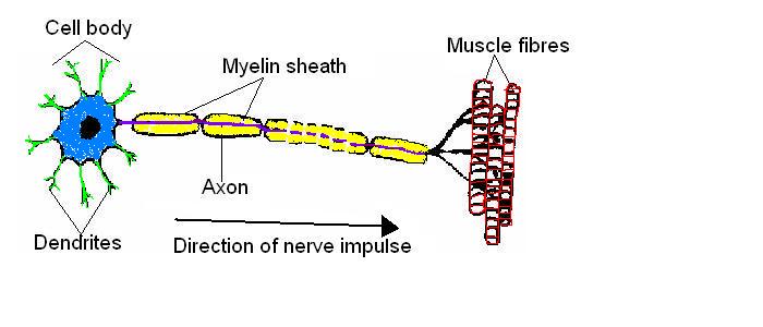
Label Neuron Anatomy Printout - EnchantedLearning.com Read the definitions, then label the neuron diagram below. axon - the long extension of a neuron that carries nerve impulses away from the body of the cell. cell body - the cell body of the neuron; it contains the nucleus (also called the soma) dendrites - the branching structure of a neuron that receives messages (attached to the cell body)

Neuron diagram labeled
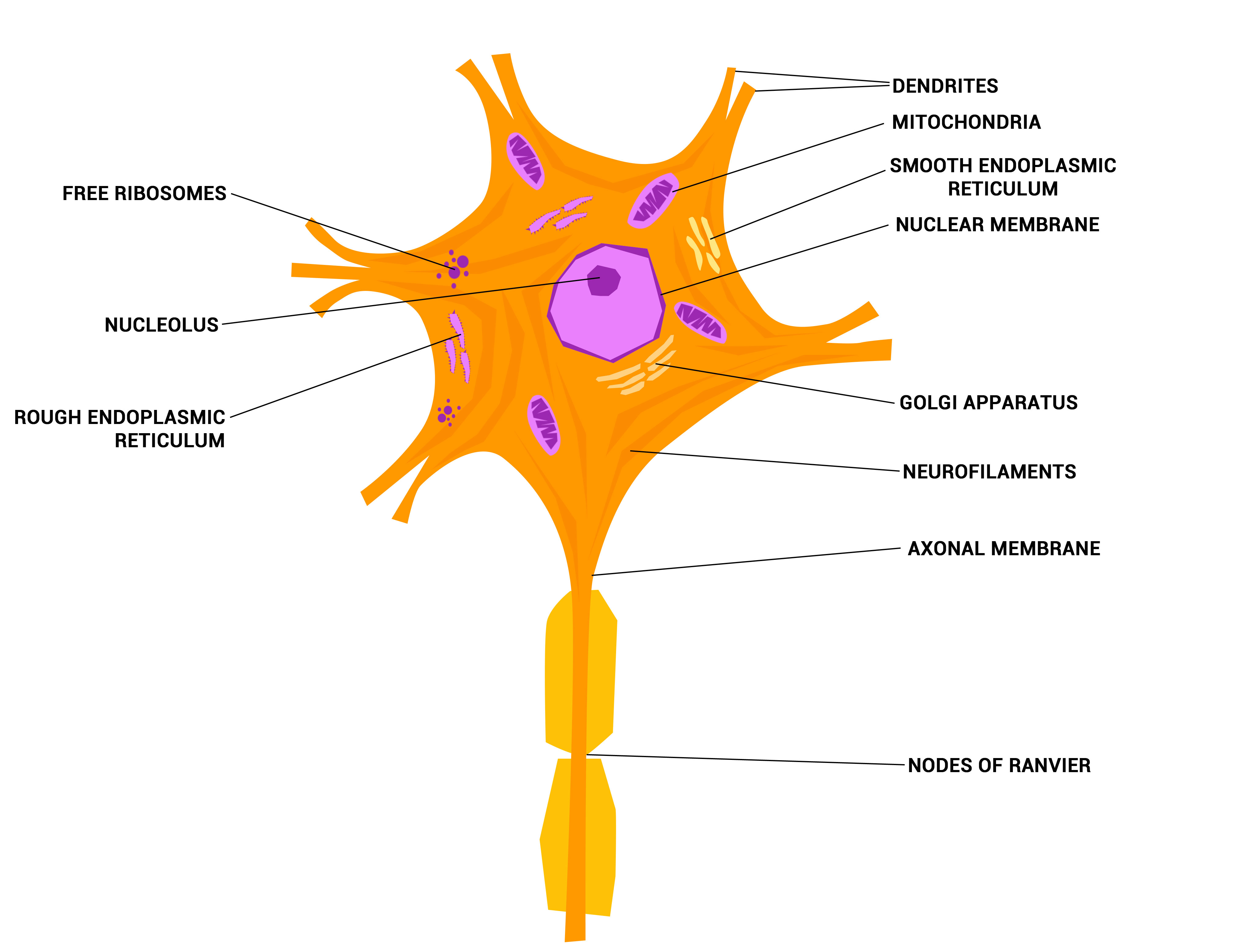
A Labelled Diagram Of Neuron with Detailed Explanations Diagram Of Neuron A neuron is a specialized cell, primarily involved in transmitting information through electrical and chemical signals. They are found in the brain, spinal cord and the peripheral nerves. A neuron is also known as the nerve cell. Labeled Diagram Of A Neuron Illustrations, Royalty-Free Vector Graphics ... Browse 20 labeled diagram of a neuron stock illustrations and vector graphics available royalty-free, or start a new search to explore more great stock images and vector art. Newest results Reflex ARC sensory neuron pathway from stimulus to response... Nerve cell with magnifying glass line icon. Neural tissue... The nervous system Neurons (With Diagram) - Biology Discussion A neuron consists of main cell body and cytoplasmic processes arising from it. ADVERTISEMENTS: (i) Cell body (= Cyton or Soma): It varies in size and form. It may be up to 13.5 µm in diameter and may be irregular, spherical, oval, rounded, star-shaped or pyramidal. Like a typical cell it consists of cytoplasm, nucleus and cell membrane.
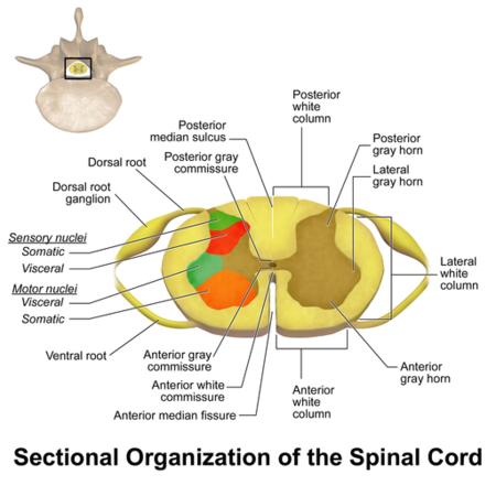
Neuron diagram labeled. Overview of neuron structure and function - Khan Academy Anatomy of a neuron. Neurons, like other cells, have a cell body (called the soma ). The nucleus of the neuron is found in the soma. Neurons need to produce a lot of proteins, and most neuronal proteins are synthesized in the soma as well. Various processes (appendages or protrusions) extend from the cell body. Neuron Labeling - Anatomy and Physiology Diagram | Quizlet Start studying Neuron Labeling - Anatomy and Physiology. Learn vocabulary, terms, and more with flashcards, games, and other study tools. Labeled Neuron Diagram | Science Trends Neurons are a type of cell and are the fundamental constituents of the nervous system and brain. Neurons take in stimuli and convert them to electrical and chemical signals that are sent to our brain. There are 3 major kinds of neurons in the spinal cord: sensory, motor, and interneurons. Neurons communicate vie electrical signals produced by ... Neuron Diagram Unlabeled - Wiring Diagram Pictures Find nerve cell diagram Stock Images in HD and millions of other royalty-free stock Related: axon and dendrites, neuron myelin, cell education, neural cells, . Read the definitions, then label the neuron diagram below. axon - the long extension of a neuron that carries nerve impulses away from the body of the cell.
Neuron (Nerve Cell) Types, Structure and Function Anatomy of a Neuron. The neuron contains the soma (cell body) from which extend the axon (a nerve fiber conducting electrical impulses away from the soma) and dendrites (tree-like structures that receive signals from other neurons). ... A relay neuron (also known as an interneuron) allows sensory and motor neurons to communicate with each other ... File:Complete neuron cell diagram en.svg - Wikipedia English: Complete neuron cell diagram. Neurons (also known as neurones and nerve cells) are electrically excitable cells in the nervous system that process and transmit information. In vertebrate animals, neurons are the core components of the brain, spinal cord and peripheral nerves. Own work. PDF Neurotransmission Fact Sheet - NIDA.NIH.GOV 3. The space between the dendrites of one neuron and the axon of another neuron is called the synapse. 4. The nucleus of a neuron is where genetic material is stored. 5. Neurons that send information from sensory organs, such as the skin or eyes, to the central nervous system are called sensory (or afferent) neurons. 6. A Labelled Diagram of Neuron with Detailed decription A neuron is a type of cell that is largely responsible for conveying information via electrical and chemical impulses. The brain, spinal cord, and peripheral nerves all contain them. The nerve cell is another name for a neuron. The structure of a neuron changes depending on its form and size, as well as its function and location.
Neuroglia Diagram - schematron.org Glial Cells (neuroglia) are the non-excitable supporting cells of the nervous system. All glial cells are much smaller but far more numerous than the nerve cells.Neuroglia. A multi-part image shows a micrograph of a neuron and six types of neuroglia cells. The micrograph of the neuron shows several tiny dot-like structures labeled neuroglia. Neuron - Label - Labelled diagram Neuron - Label. Share Share by Aspillane. Y12 Y13 Psychology. Like. Edit Content. Embed. More. Leaderboard. Show more Show less . This leaderboard is currently private. Click Share to make it public. This leaderboard has been disabled by the resource owner. This leaderboard is disabled as your options are different to the resource owner. ... Neuron Diagram, Structure & Function | What Is a Neuron? - Video ... A detailed diagram of a neuron, showing the synapse, cell body, axon, and dendrites. Dendrites One unique feature of neurons is that they have extensions known as dendrites that extend outward from... Nervous System - Label the Neuron Nervous System - Label the Neuron Nervous System - Neuron: Nerve Cell Name: Choose the correct names for the parts of the neuron. (1) (2) (3) (4) (5) (6) This neuron part receives messages from other neurons. (7) This neuron part sends on messages to other neurons. (8) This neuron part gives messages to muscle tissue. (9)
Nervous System Worksheet Answers - WikiEducator
Neuron under Microscope with Labeled Diagram - AnatomyLearner The neuron structure has two main components: the cell body and the neuron processes (axons and dendrite). Let's see the neuron histology slide labelled diagram and try to find out the below-mentioned characteristics - Presence of an identifiable cell body (soma) that locates in the brain's grey matter (according to the slide image).

Labeled illustration depicting normal neuron synapse activity Stock ...
What Is a Neuron? - Definition, Structure, Parts and Function There are three different types of neurons: Sensory Neurons The sensory neurons convert signals from the external environment into corresponding internal stimuli. The sensory inputs activate the sensory neurons and carry sensory information to the brain and spinal cord. They are pseudounipolar in structure. Motor Neurons
Neurons - The crazy wires in our body. – Doctor Jana
Sensory Neuron Diagram Stock Illustrations - Dreamstime Download 389 Sensory Neuron Diagram Stock Illustrations, Vectors & Clipart for FREE or amazingly low rates! New users enjoy 60% OFF. 186,471,427 stock photos online. Stock Photos; ... Neuron. Labeled diagram of the Neuron, nerve cell that is the main part of the nervous system. Abstract grey mesh background.

Pin on biology
Unlabeled Neuron Diagram Rlawson ×× ( bytes) Unlabeled diagram of a motor neuron by Ruth Lawson, Otago Polytechnic. Neuron Anatomy Activity. The parts of the neuron have been labeled. Your challenge is to write the correct name for each part and explain what it does. An interactive quiz covering General Structure of a Neuron through multiple- choice questions and ...

Structure Of A Typical Neuron Stock Illustration - Download Image Now ...
A Guide to Understand Neuron with Neuron Diagram | EdrawMax Online To properly understand the coordination between the brain and the body, the students must learn about the neurons. They can use neuron-labeled diagrams while learning the complex structure of neurons. Creating a neuron-labeled image by hand can be difficult. The students must use the EdrawMax Online tool to make a high-quality neuron diagram . 2.

Print The Nervous System flashcards | Easy Notecards
Neuron Diagram Labeled | EdrawMax Template It is an effective form of self-assessment, enabling students to check their understanding. In the following diagram, we have illustrated the important parts of the Neuron. In the following Neuron labeled diagram, we have dendrite, cell body, axon, myelin sheath, Schwann cell, a node of Ranvier, axon terminal, and nucleus.

Axon - Structure and Functions
Diagram Quiz on Neuron Structure and Function (Labeling Quiz) Diagram Quiz on DNA replication. 1. Identify the cell type in the above figure. 2. In the figure, labeled '1' receives impulses from adjacent neuron. It is called the. 3. In the figure, labeled '2' is the short filaments from the cell body that carries impulses from dendrites to the cell body which is the. 4.
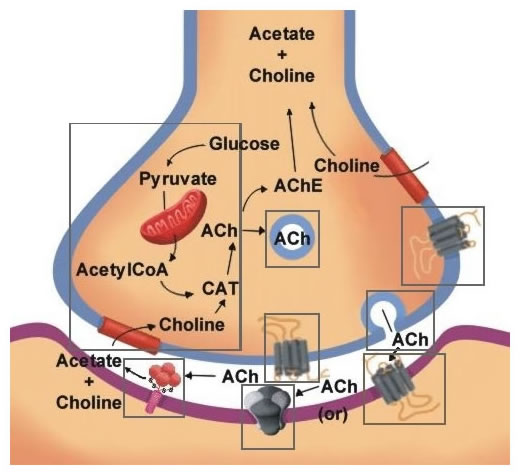
Acetylcholine Neurotransmission (Section 1, Chapter 11) Neuroscience ...
how to draw structure of neuron/neuron diagram labelled ... - YouTube Please watch: "cell structure and functions / animal cell vs plant cell / parts of cell / ch 8 science class 8 cbse" ...
Ganglion - Physiopedia
Neuron Diagram & Types | Ask A Biologist They pass signals from one neuron to the next inside the central nervous system. Pyramidal neurons are named after the shape of their cell body, which looks like a pyramid. They have one axon and two main dendrite branches. These cells pass signals inside the brain and tell your muscles to move.
Post a Comment for "40 neuron diagram labeled"