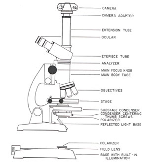
40 diagram of compound microscope with labelling
Parts of a microscope with functions and labeled diagram Q. List down the 18 parts of a Microscope. 1. Ocular Lens (Eye Piece) 2. Diopter Adjustment 3. Head 4. Nose Piece 5. Objective Lens 6. Arm (Carrying Handle) 7. Mechanical Stage 8. Stage Clip 9. Aperture 10. Diaphragm 11. Condenser 12. Coarse Adjustment 13. Fine Adjustment 14. Illuminator (Light Source) 15. Stage Controls 16. Base 17. (b) Why both objective and eyepiece of a compound microscope must have ... (a) Draw the labelled ray diagram for the formation of image by a compound microscope. Derive an expression for its total magnification (or magnifying power), when the final image is formed at the near point. (b) Why both objective and eyepiece of a compound microscope must have short focal lengths?
Compound Microscope Parts, Functions, and Labeled Diagram So, a compound microscope with a 10x eyepiece magnification looking through the 40x objective lens has a total magnification of 400x (10 x 40). Specimen or slide: The object used to hold the specimen in place along with slide covers for viewing. ... Functions, and Labeled Diagram. Posted by Fred Koenig on Nov 18th 2020. Compound Microscope ...
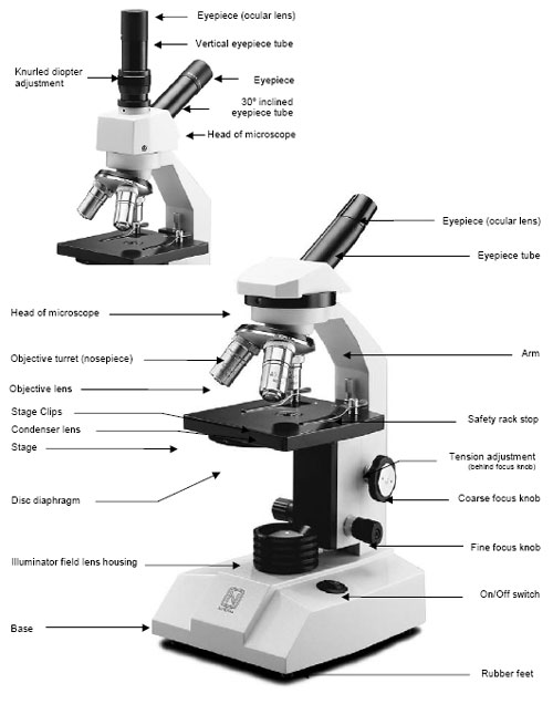
Diagram of compound microscope with labelling
Labeling the Parts of the Microscope Labeling the Parts of the Microscope This activity has been designed for use in homes and schools. Each microscope layout (both blank and the version with answers) are available as PDF downloads. You can view a more in-depth review of each part of the microscope here. Download the Label the Parts of the Microscope PDF printable version here. BYJUS BYJUS A Study of the Microscope and its Functions With a Labeled Diagram A Study of the Microscope and its Functions With a Labeled Diagram To better understand the structure and function of a microscope, we need to take a look at the labeled microscope diagrams of the compound and electron microscope. These diagrams clearly explain the functioning of the microscopes along with their respective parts.
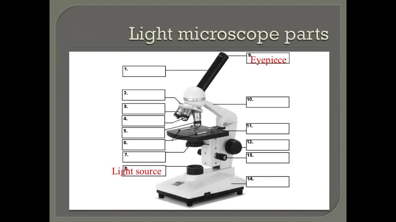
Diagram of compound microscope with labelling. Compound microscope - their parts and function - Microscopy4kids Labeled diagram of a compound microscope. Optical components of a compound microscope. The term "compound" refers to the microscope having more than one lens. Compound microscopes generate magnified images through an aligned pair of the objective lens and the ocular lens. In contrast, "simple microscopes" have only one convex lens and ... Labelled Diagram of Compound Microscope - Biology Discussion The below mentioned article provides a labelled diagram of compound microscope. Part # 1. The Stand: The stand is made up of a heavy foot which carries a curved inclinable limb or arm bearing the body tube. The foot is generally horse shoe-shaped structure (Fig. 2) which rests on table top or any other surface on which the microscope in kept. Labeling the Parts of the Microscope | Microscope activity, Science ... Description Worksheet identifying the parts of the compound light microscope. Answer key: 1. Body tube 2. Revolving nosepiece 3. Low power objective 4. Medium power objective 5. High power objective 6. Stage clips 7. Diaphragm 8. Light source 9. Eyepiece 10. Arm 11. Stage 12. Coarse adjustment knob 13. Fine adjustment knob 14. Base Parts of the Microscope with Labeling (also Free Printouts) 5. Knobs (fine and coarse) By adjusting the knob, you can adjust the focus of the microscope. The majority of the microscope models today have the knobs mounted on the same part of the device. Image 5: The circled parts of the microscope are the fine and coarse adjustment knobs. Picture Source: bp.blogspot.com.
(i) Draw a neat labelled ray diagram of a compound microscope. Explain ... The eyepiece forms its image A'' B'' which is virtual, erect and magnified. Thus the final image A'' B'' formed by the microscope is inverted and magnified and its position is outside the objective and eyepiece towards objective lens. Magnifying power of compound microscope is. for final image at distance of distinct vision. for final image at ... Compound Microscope- Definition, Labeled Diagram, Principle, Parts, Uses Parts of a Compound Microscope Eyepiece And Body Tube. The eyepiece is the lens through which the viewer looks to see the specimen. It usually contains a 10X or 15X power lens. The body tube connects the eyepiece to the objective lenses. Objectives and Stage Clips Objective Lenses are one of the most important parts of a Compound Microscope. Compound Microscope Parts - Labeled Diagram and their Functions - Rs ... Labeled diagram of a compound microscope Major structural parts of a compound microscope There are three major structural parts of a compound microscope. The head includes the upper part of the microscope, which houses the most critical optical components, and the eyepiece tube of the microscope. Compound Microscope: Parts of Compound Microscope - BYJUS (A) Mechanical Parts of a Compound Microscope 1. Foot or base It is a U-shaped structure and supports the entire weight of the compound microscope. 2. Pillar It is a vertical projection. This stands by resting on the base and supports the stage. 3. Arm The entire microscope is handled by a strong and curved structure known as the arm. 4. Stage
Draw a neat labelled diagram of a compound microscope and explain its ... Dividing and multiplying by I1 G1 on the right side, we get Magnifying power of the objective (m0) = I1G1/OJ = Height of the image due to the objective. Magnifying power of the eye piece (me) = IG/I1G1 = Height of the final image / Height of the object for the eyepiece. ∴ m = m0 × me ..... (1) Compound Microscope Parts, Function, & Diagram | What is a Compound ... The base of the compound light microscope is the bottom portion of the compound microscope. It functions to support the entire compound microscope. The base can be set on a table or lab bench, and... Compound Microscope Labeled Diagram | Quizlet QUESTION. The total magnification of a specimen being viewed with a 10X ocular lens and a 40X objective lens is. 15 answers. QUESTION. a mosquito beats its wings up and down 600 times per second, which you hear as a very annoying 600 Hz sound. if the air outside is 20 C, how far would a sound wave travel between wing beats. 2 answers. Microscope Parts, Function, & Labeled Diagram - slidingmotion Condenser. The condenser is to focus the light, which passes from the microscopic illuminator to the specimen. This condenser is located just below the diaphragm. This diaphragm is one of the important parts of the compound microscope which will help to get an accurate and sharp image. The condenser has a magnification power of 400X and above.
MICROSCOPE DIAGRAM - Unmasa Dalha
Parts of a Compound Microscope and Their Functions Compound microscope magnification is determined by multiplying the eyepiece and objective powers. When viewed through a 5X eyepiece with a 10X objective, an item is magnified 5 x 10=50 times. The magnification is 10 x 45 = 450 times when using a 10X eyepiece and a 45X objective. How to Use the Compound Microscope

Microscope labeling - YouTube
16 Parts of a Compound Microscope: Diagrams and Video In compound microscopes with two eye pieces there are prisms contained in the body that will also split the beam of light to enable you to view the image through both eye pieces. 2. Arm. The arm of the microscope is another structural piece. The arm connects the base of the microscope to the head/body of the microscope.
How to properly use a compound light microscope - YouTube
Diagram of a Compound Microscope - Biology Discussion The size of objects viewed under the compound microscope can be accurately determined using a micrometer. The latter consists of two scales, the eyepiece scale, (also called 'graticule' or 'ocular') and the stage micrometer scale. The eyepiece scale is calibrated with the help of stage micrometer and the former is then used for measurements.
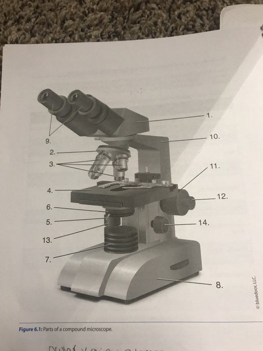
Diagram Compound Microscope Parts And Functions - Micropedia
Microscope Types (with labeled diagrams) and Functions Compound microscope labeled diagram Compound microscope functions: It finds great application in areas of pathology, pedology, forensics etc Its greater order of magnification allows for deeper study of microbial organisms to Detect the cause of diseases Study the mineral composition in soils
December - Mrs. Beltz's Biology Class
Microscope Labeling Diagram | Quizlet Animal Cell Labeling 1. 9 terms. PGFry210. Plant Cell Labeling 1. 10 terms. PGFry210. Animal Cell Labeling 2 ... Parts of a Microscope (Bio Quiz) 13 terms. angela2sweet. An Introduction to the Compound Microscope. 28 terms. MiracleFaith98. OTHER SETS BY THIS CREATOR. Unit 2 Lesson 7 - Biotechnology ... Diagrams. Flashcards. Mobile. Help. Sign ...

Choosing a Microscope | Make: DIY Projects and Ideas for Makers
Compound Microscope - Diagram (Parts labelled), Principle and Uses See: Labeled Diagram showing differences between compound and simple microscope parts Structural Components The three structural components include 1. Head This is the upper part of the microscope that houses the optical parts 2. Arm This part connects the head with the base and provides stability to the microscope.

Compound Microscope Labeled - ClipArt Best
Microscope Labeling - The Biology Corner Students label the parts of the microscope in this photo of a basic laboratory light microscope. Can be used for practice or as a quiz. ... Microscope Labeling . Microscope Use: 15. When focusing a specimen, you should always start with the _____ objective. 16. When using the high power objective, only the _____ knob should be used. 17. The ...

Diagram Of Microscope Example Binocular And Their Functions Compound ...
Microscope Drawing And Label - Painting Valley All the best Microscope Drawing And Label 33+ collected on this page. Feel free to explore, study and enjoy paintings with PaintingValley.com ... label; microscope; diagram; compound; parts; light; labeling; functions; microscopic; blank; labeled; biology; microscopy; labelled; beautiful; Like JPG. Compound Microscope ... 496x600 35 0. Like JPG ...

All Saints Online: Diagram for Labelling: Microscope
Draw a labelled diagram of an image formed by a compound microscope ... Click here👆to get an answer to your question ️ Draw a labelled diagram of an image formed by a compound microscope, with the image at least distance of distinct vision. Write any one expression for its magnifying power. Solve Study Textbooks Guides. ... Draw a labelled ray diagram of an image formed by a compound microscope, when the final ...
Molecular Expressions: Microscopy Publications - Fascinating Photos ...
Label the microscope — Science Learning Hub All microscopes share features in common. In this interactive, you can label the different parts of a microscope. Use this with the Microscope parts activity to help students identify and label the main parts of a microscope and then describe their functions. Drag and drop the text labels onto the microscope diagram.

Compound Microscope Diagram - Micropedia
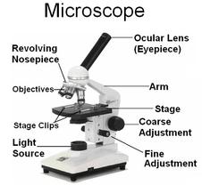
Microscope Parts and Functions With Labeled Diagram and Functions How ... Coarse adjustment: Brings the specimen into general focus. Fine adjustment: Fine tunes the focus and increases the detail of the specimen. Nosepiece: A rotating turret that houses the objective lenses. The viewer spins the nosepiece to select different objective lenses. Objective lenses: One of the most important parts of a compound microscope ...
Post a Comment for "40 diagram of compound microscope with labelling"